Advertisement
Advertisement
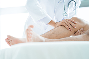
Hip
Avascular Necrosis (Osteonecrosis) of the Hip
Bursitis of the Hip (Trochanteric Bursitis)
Core Decompression for Avascular Necrosis of the Hip
Femoral Nerve Block
Femoroacetabular Impingement (FAI)
Femur Fracture Fixation (Stryker® Gamma Nail)
Femur Fracture Fixation (Dynamic Hip Screw Method)
Femur Fracture Fixation with Intramedullary Rod
Femur Fractures
Hip Arthroscopy
Hip Dislocation
Hip Fracture
Hip Fracture
Hip Fracture Prevention
Femur Fracture Fixation (Surgical Screws Method)
Hemiarthroplasty (Bipolar Prosthesis)
Hip Joint Injection (Ultrasound or Fluoroscopic Method)
Internal Screw Fixation for Slipped Capital Femoral Epiphysis (SCFE)
Hip Joint Injection
Labral Tear of the Hip (Acetabular Labrum Tear)
Total Hip Replacement (Minimally-Invasive Method, Large-Diameter Bearing)
Loose Bodies in the Hip
Muscle Strain Injuries of the Hip
Osteonecrosis of the Hip
Partial Hip Resurfacing (Wright)
Periacetabular Osteotomy (PAO)
Legg-Calve-Perthes Disease (LCPD)
PRP Therapy for Hip Arthritis
PRP Therapy for Hip Arthritis (AcCELLerated Biologics)
Sacroiliac Joint Fusion (Rialto™ SI Fusion System)
Slipped Capital Femoral Epiphysis (SCFE)
Snapping Hip
Stem Cell Therapy for Avascular Necrosis of the Hip
Stem Cell Therapy for Hip Pain
Femoroacetabular Impingement Surgery (Open Method)
Hip Resurfacing
Total Hip Resurfacing (Wright)
Transient Osteoporosis of the Hip
Focus On: Slipped Capital Femoral Epiphysis
Sex Positions After Joint Replacement
Hip Arthroscopy: Removing Loose Bodies
Hip Arthroscopy- Labral Tears
Hip Arthroscopy: Repairing Chondral Damage
Hip Arthroscopy: Repairing Femoroacetabular Impingement
Hip Arthroscopy: Repairing Synovitis and Arthritis
Hip Arthroscopy: Before Surgery
Hip Arthroscopy: After Surgery
How Your Hip Works
After Hip Surgery- Mastering Daily Tasks
After Hip Surgery- Getting Dressed
Hip Safety: Getting Into and Out of Bed
After Hip Surgery- Getting Into and Out of a Car
Hip Precautions
Hip Safety: Sleeping Positions
After Hip Replacement- Sitting Safely
Hip Safety: Using the Toilet
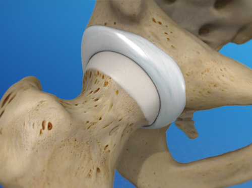
Total Hip Replacement
Understanding Hip Replacement
After Hip Replacement: Recovering in the Hospital
After Hip Replacement: Continuing with Hospital Recovery
After Total Hip Replacement: Returning to Activity
After Hip Replacement: Home Safety
After Total Hip Replacement: Recovering at Home
Getting Ready for Hip Replacement Surgery
Before Total Hip Replacement: Your Conditioning Program
Before Total Hip Replacement: Preparing for Your Recovery
Pre-Hip Replacement: Ankle Pumps, Quad Sets, Leg Raises
Pre-Hip Replacement Exercises for the Legs
Exercises Before Hip Replacement: To Help with Walker or Crutch Use
Discharge Instructions for Total Hip Replacement Surgery
After Hip Replacement: Managing Your Pain
After Hip Replacement: When to Call Your Surgeon
Understanding Hip Resurfacing
Understanding Minimally Invasive Total Hip Replacement
Having Hip Resurfacing
Treatment for Iliotibial (IT) Band Syndrome
Having Minimally Invasive Total Hip Replacement
Understanding Trochanteric Bursitis
Step-by-Step: Using Log-Roll to Get into Bed (Hip Care)
Step-by-Step: Using Log-Roll to Get Out of Bed (Hip Care)
Core Decompression of the Hip
Femoroacetabular Impingement
Knee
Anterior Cruciate Ligament (ACL) Tear in Children: Care Instructions